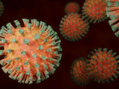
Human Papilloma Virus

Abstract
Numbers of new cervical cancer cases are increasing constantly although this tumor is one of the best preventable malignancies of all relevant human cancers. We present the case of a female patient, whose Papanicolaou test detected HPV infection and the cervical and vulvar biopsy showed carcinoma of the cervix uteri and vulva. First, she made the vaccination with Gardasil 9 and after it was made the total hysterectomy. She recovered after the surgical intervention and she make periodic medical examination.
Table of Contents:
1. Introduction
2. Results and discussion
3. Discussions
4. Conclusions
1. Introduction
Human Papilloma Virus (HPV) infection is found in up to half of vulvar cancers. Almost all HPV infections are transmitted during sexual contact. The most common cancer-producing viruses are HPV types 16, 18, and 33 [1].
Cervical cancer is the third most common cancer in woman worldwide. Is a disease that develops quite slowly and begins with a precancerous condition known as dysplasia. Dysplasia is easily detected in a routine Pap smear. Infection with the common papillomavirus is a cause of approximately 90% of all cervical cancers [2, 3].
2. Results and discussion
The case study involved a 50-year-old female patient reported with genital problems in the IInd Department of Obstetrics and Gynecology at the “Pius Brânzeu” County Emergency Hospital in Timisoara. Her past medical history is notable for hypertension. She takes no medications. She is married with two children. Her first sexual contact was at age 16, and she has had four partners in her lifetime. She has been smoking 20 cigarettes per day for the last 25 years. She reported abnormal bleeding after sexual intercourse and between periods, unpleasant vaginal discharge, pain during sex, lower back pain, persistent itchiness, soreness and burning within the vulvar space and an ulceration on vulvar space that persist for over 2 months.
A gynecologic examination revealed obvious tumor growth confined to her cervix with no signs of extension to her vagina and a vulvar tumor. The Papanicolaou test detected the koilocit cell type that are suggestive for a genital HPV infection. Human papillomavirus DNA typing revealed the presence of human papillomavirus-16.
It was recommended a cervical and vulvar biopsy. The cervical and vulvar biopsy showed moderately differentiated (tumor grade II) large-cell keratinizing squamous cell carcinoma of the cervix uteri and vulva. Neither lymphatic nor venous vascular space involvement was reported, but dense inflammatory cell infiltration of the tumor stroma was noted. Clinical staging was completed by cystoscopy, proctoscopy, and chest radiography (as allowed for accurate clinical staging by the International Federation of Gynecology and Obstetrics [FIGO]), which revealed stage IB2 cancer.
As a method of treatment was decided HPV vaccination with Gardasil 9. After vaccination the immune system (the body’s natural defense system) stimulate the formation of antibodies against the new HPV types in the vaccine to help protect against diseases caused by these viral strains. After the vaccine both the vulvar and cervical tumors decreased in size.
A radical hysterectomy with bilateral salpingo-oophorectomy was made after 3 months of the vaccine. During the surgery everything went in the physiological parameters. The patient recovered quickly after surgery intervention and now she make periodic medical examination.
3. Discussions
Human papillomaviruses encompass more than 120 different types that may infect human skin and mucosa. Only 13-15 of these are found in cervical cancers and other malignancies and are called ‘high risk’ HPV (HPV-HR). HPV 16 is the most important HPV-HR-type; it is linked to approximately 50% of cervical cancers worldwide. HPV 18 ranks second, HPV 16 and 18 are associated with two thirds of all cervical cancers as well as subsets of cancers of the vulva, vagina, penis, anus, oropharynx and skin [4]. In our case report, unfortunately Human papillomavirus DNA typing revealed the presence of human papillomavirus-16.
Squamous cell carcinoma is far more common than verrucous carcinoma in the vulva. The clinical and morphologic distinctions between these neoplasms are important to understand because of their contrasting biologic behavior and treatment. Both cancers present with symptoms of pruritus and a noticeable mass. On examination, both tumors commonly occur on the labia and are exophytic. If infection occurs in association with verrucous carcinoma, the resulting induration of the surrounding tissue as well as reactive regional lymph node enlargement may fool the clinician into making an erroneous diagnosis of advanced squamous cell carcinoma [5].
The curative treatment of vulvar cancer requires consideration of both the primary focus of disease in the vulva and the inguinal lymph nodes, which are the regional lymph nodes of the vulva. The cardiovascular system must be explored carefully before surgery [6, 7, 8, 9].
In current treatment, surgery is the first choice, and historically there has been a transition away from radiation therapy toward surgical procedures [10, 11].
According to the American College of Radiology (ACR) Appropriateness Criteria, periodic follow-up is recommended once every 3 months for the first 2 years after treatment, followed by periodic observation at longer intervals thereafter. However, the grounds for these recommendations are not explained. Some retrospective studies of multiple cases showed 70-80% of relapsing cases occur within the first 2 years after initial treatment. The recurrence rate declines for the third and subsequent years, but recurrences have been confirmed even after 5 years [12].
Most cases in the old VIN 1 category are LSIL, and doubt exists as to their significance as neoplastic lesions. However, the old histopathological definition of VIN 1 includes dVIN which, unlike LSIL, is a neoplastic lesion and must be separated from VIN 1 even though the frequency of dVIN occurrence is very low [13]. On that understanding, it is preferable to avoid invasive treatment for LSIL and to follow-up periodically. On the other hand, HSIL and dVIN are neoplastic lesions which require treatment. In a systematic review of the literature, it was found that 9% of untreated cases of VIN 3 progressed to invasive carcinomas, and 3% of cases that are surgically excised had occult invasive carcinoma [14], indicating that biopsy under colposcopy is important to exclude invasion [15]. Clinically the evidence for a clear distinction between VIN 2 and VIN 3 has not been demonstrated, and HSIL, which includes both VIN 2 and VIN 3, must be managed with the same due consideration [16]. Because HSIL is caused by HPV infection, multiple foci of disease can appear in a wide-ranging area of the vulva and can also appear, simultaneously or allochronically, in and around the uterine cervix, vagina and anus. Careful examination of all of these areas is required.
In the management of these cases it is important to think at the age of the patient and at the patient’s desire to have a baby in the future [17,18, 19].
4. Conclusions
Cervical sCreening is one of the best defenses against the development of cervical cancer. Clinicians should make a point of routinely enquiring about the date and result of the patient’s last cervical smear test. This could provide an important opportunity for health education, as many older women are still under the misconception that cervical cancer is a disease that only affects young, promiscuous women.Cervical cancer is highly preventable through HPV vaccination and treatment of pre-clinical lesions detected by screening.
Contributo selezionato da Filodiritto tra quelli pubblicati nei Proceedings “4th National Congress of HPV - 1st Congress of the Society of Endometriosis and East-European Infertility - 2018”
Per acquistare i Proceedings clicca qui.
Contribution selected by Filodiritto among those published in the Proceedings “4th National Congress of HPV - 1st Congress of the Society of Endometriosis and East-European Infertility - 2018”
To buy the Proceedings click here.
MITRACHE Dana [1]
ILINA Razvan [2]
VRINCEANU Luminita [3]
BRISAN Cosmin [1]
[1] “Pius Brinzeu” Emergency Clinic County Hospital Timisoara, IInd Department of Obstetrics and Gynecology, Timisoara (ROMANIA)
[2] “Victor Babes” University of Medicine and Pharmacy Timisoara, Department of Surgery, Timisoara (ROMANIA)
[3] Medline, Tirgu Jiu (ROMANIA)


