
External approach to frontal sinus


Abstract
In this study we report our experience in external approach for frontal sinus. A retrospective review was performed of frontal sinus procedures in our clinic from 2015 to 2018. The goal was to evaluated the patients and the results of an external approach for frontal sinus inflammatory disease and the possible ophthalmologic affections.
Table of Contents:
1. Introduction
2. Methodology
3. Results
4. Discussion
5. Conclusions
1. Introduction
The radical approach to frontal sinus is the surgical procedure where the sinus passes through the frontal-orbital pathway, it connects along the sinus mucosa and other intrasinusal pathological formations, and a large drainage is achieved by extending the frontal-nasal passage. Several techniques for external approach have been described, which can be done alone or in combination with the endoscopic approach [1]. The external approach of the frontal sinus is Bock trepanopunction, Jansen, Jaques-Durand, Killian, Kuhnt-Riedel, Claus-Matis, Ogston-Luc, Ledoux, Ogston-Taptas I and II, Lynch, Neal-Lake and Sewall-Boyden procedures, osteoplastic, reconstructive and obliterative procedure. Emergency indications of the external approach may be sinusitis, acute suppurative relapses sinusitis or bone, meningoencephalic or orbital complications [2].
Preoperative assessment of patients was greatly improved by the large-scale introduction of computed tomography, magnetic resonance imaging and endoscopic nasal examination, the last one assessing particularly the ostiomeatal complex [3].
2. Methodology
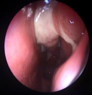
Between 2015-2018, in our ENT Department from Timisoara 15 patients were operated by the frontal sinus external approach. Six patients (40%) presented osteomas, 5 patients (33.33%) frontal sinusitis, with persistent headache, 3 patients (20%) mucoceles and 1 patient (6.66%) was diagnosed with a nasopharyngeal tumour (estezioneuroblastoma std. III). The gender distribution included 7 women (46.66) and 8 males (53.33%). Distribution by age group was as follows: 10-20 years (1 case); 20-30 years (8 cases); 30-40 years (3 cases); 40-50 years (2 cases); 50-60 years (1 case). The external approach of the frontal sinus was performed for acute recurrent sinusitis or for orbital complications. The diagnosis was based on symptomatology (frontal headache, nasal obstruction and discharge, orbital pain), ENT and ophthalmologic clinical examination, nasal endoscopy (Fig. 1), CT and MRI.
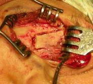
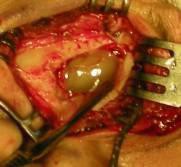
The Ogston-Luc procedure (Fig. 2 and 3) was performed in most of the cases (14 patients). The anterior craniofacial approach associated with the frontal sinus external approach was performed in case of malignant tumours (1 case). The drainage tube has been kept for 21 days. From ophthalmologic point of view the patients presented periorbital tenderness (5 patients, 33%), swelling (4 patients, 26,6%), chemosis (4 patients, 26,6%), decreased visual acuity (3 patients, 20%) and restriction of extraocular movements (1 patient, 6,66%).
3. Results
The Ogston-Luc procedure was performed in the 15 patients, in 12 patients (80%) the frontal sinus anterior wall was restored. Intra and postoperative patient’s evolution was favourable, and postoperative monitoring was performed by nasal endoscopy.
Post-operative follow-up did not reveal any ENT or ophthalmologic complications: immediate (loss of cerebrospinal fluid, meningitis, wound infection), tardive (mucus, recurrent sinusitis). The symptoms were resolved in all cases. Two patients (1.33%) presented supraorbital neuralgia and other 2 patients (1.33%) presented frontal paraesthesia.
4. Discussion
Knowledge of pathophysiology as well as the surgeon’s experience are essential in establishing the surgical technique. In the literature, it is recommended to treat the endonasal pathology gradually by opening the frontal cavity (drainage procedure type I or sinusotomy), to partial drainage through the frontal sinus floor (type II), until the bilateral frontal sinuses are drained (type III or Modified Lothrop).
Ethiopathological factors should be assessed prior identification of the treatment options [4-8]. Hahn Samuel et al., performed a retrospective study on 38 patients with external approach to frontal sinus (14 patients) or the combined approach (28 patients). The major indication for surgical intervention was neo-osteogenesis of the frontal recess, most commonly associated with frontal sinus trauma, endoscopic surgery and frontal bone osteoarthritis. 28 patients (78%) were subjected to a history of sinus surgery [9].
Alexander G. Chiu confirms that most surgeons agree that the optimal surgical procedure is that which causes the disappearance of symptoms and sinus frontal infection, with the absence of orbital or skull base complications. Ideal surgery also ensures minimal morbidity with a minimal aesthetic defect [10].
Due to the orbital complications a mixed team formed by an ENT surgeon and an ophthalmologist assessed all the cases [11-23]. The nasopharyngeal tumour (estezioneuroblastoma std. III) was biopsied prior removal. Accurate assessment of the head and neck tumours is a very important step [24].
An optimal surgical technique should have the following features: be applied only once; to lead to the disappearance of the symptoms and disease, but with the maintenance of sinus function and region minimal deformation.
5. Conclusions
The frontal sinus approach can be performed endoscopically, through external approach techniques or by a combined approach. Choosing a technique depends mainly on the extent of the disease, the local anatomy, the type of previous surgery (if it exists), and last but not least on the surgeon’s experience.
The success of any procedure is based on resolving the symptoms, eradicating the infection and preserving the aesthetic aspect.
The most common orbital complications encountered were periorbital tenderness, swelling, chemosis, decreased visual acuity and restriction of extraocular movements.
The Authors:
BALICA Nicolae Constantin [1]
POENARU Marioara [1]
DOROS Caius [1]
HORHAT Delia Ioana [1]
BOIA Eugen Radu [1]
DUMITRU Cristina Stefania [1]
[1] ENT Department, “Victor Babes” University of Medicine and Pharmacy Timisoara (ROMANIA)
Contributo selezionato da Filodiritto tra quelli pubblicati nei Proceedings “11th Conference of Ophthalmology with International Participation - 2019”
Per acquistare i Proceedings clicca qui.
Contribution selected by Filodiritto among those published in the Proceedings “11th Conference of Ophthalmology with International Participation - 2019”
To buy the Proceedings click here.
